


Screening results for full-term neonatal eye diseases from 2016 to 2023
A total of 17,378 full-term neonates participated in eye disease screening. Among them, 9,178 were male (52.81%), and 8,200 were female (47.19%) (Table 1). Vaginal deliveries accounted for 9,988 cases (57.47%), while cesarean sections accounted for 7,390 cases (42.53%). Initial screening identified 11,392 normal cases, with a pass rate of 65.55%, and 5,986 abnormal cases, resulting in an abnormality rate of 34.45%. Among the abnormalities, the five most common conditions were: Retinal hemorrhage, (Fig. 1a) (3,274 cases, 18.84%), Retinal white spots, (Fig. 1b) (1,440 cases, 8.29%), Retinal exudates, (Fig. 1c) (943 cases, 4.53%), Retinal pigment abnormalities, (Fig. 1d)(437 cases, 2.51%), FEVR-like lesions, (Fig. 1e) (228 cases, 1.31%). Among the 3,274 cases with retinal hemorrhages, 489 involved macular hemorrhages, including 287 unilateral cases and 202 bilateral cases.Foveal hemorrhage was identified in 45 of the 489 cases with macular hemorrhage.This study also identified 18 cases of congenital cataracts and 275 cases of other ocular abnormalities. Excluding subconjunctival hemorrhages, 141 cases of relatively rare retinal abnormalities were observed, including: choroidal coloboma, (Fig. 1f) (n = 45),vitreous opacity (n = 29),tortuous retinal arteries without other retinal changes (n = 19),peripheral retinal avascular zone (1–2 disc diameters) without other retinal changes (n = 18),corneal opacity (n = 5),abnormal cup-to-disc ratio (n = 5),persistent pupillary membrane (n = 3),eyelid hemangioma (n = 3),persistent hyaloid artery remnant, (Fig. 1g) (n = 2),abnormal retinal arteriovenous anastomosis (n = 2),microphthalmos with microcornea (n = 2),blepharophimosis (n = 2),retinoblastoma (RB), (Fig. 1h) (n = 1),persistent fetal vasculature (PFV), (Fig. 1i)(n = 1),lenticonus (n = 1),optic disc drusen (n = 1),conjunctival polyp (n = 1),and vitreous hemorrhage (n = 1).

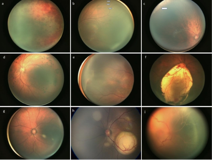
Abnormalities in Eye Disease Screening among full-term neonatal a:Retinal hemorrhage. b:Retinal white spots (several white spots in the superotemporal retina, as indicated by the arrows). c:Retinal Exudates, (Grayish-white exudative lesions in the temporal peripheral retina, as indicated by the arrows). d:Retinal pigment abnormalities,(Congenital Hypertrophy of the Retinal Pigment Epithelium, CHRPE). e:Familial exudative vitreoretinopathy (FEVR)-like fundus changes. f:Choroidal coloboma (a large coloboma inferior to the optic disc in the right eye, accompanied by minor retinal hemorrhage). g:Persistent Hyaloid Artery. h:retinoblastoma (RB). i:persistent fetal vasculature (PFV).
Screening results for full-term neonates by gender and delivery method from 2016 to 2023
During the study, ocular screening was performed on 9,178 male and 8,200 female full-term neonates. The screening results were normal in 5,984 (65.20%) males and 5,409 (65.96%) females, and abnormal in 3,194 (34.80%) males and 2,791 (34.04%) females. No significant difference in the abnormality rate was observed between male and female neonates (χ² = 1.120, P = 0.290; Table 2). A total of 9,988 vaginally delivered full-term neonates were screened, with 5,581 normal (55.88%) and 4,407 abnormal (44.12%). For 7,390 neonates delivered via cesarean section, 5,812 were normal (78.65%) and 1,578 were abnormal (21.35%). The proportion of eye abnormalities was significantly higher in vaginal deliveries than in cesarean sections (X2 = 975.299, P = 0.000, Table 2).
Retinal abnormalities in full-term neonates by delivery method from 2016 to 2023
A comparison of retinal hemorrhage, retinal white spots, and retinal exudates by delivery method is shown in Table 3. Among the 9,988 neonates delivered vaginally, 7,013 (60.25%) had no retinal hemorrhage, while 2,975 (29.79%) had retinal hemorrhage. Among the 7,390 neonates delivered via cesarean section, 7,091 (95.95%) had no retinal hemorrhage, while 299 (4.05%) had retinal hemorrhage. Retinal hemorrhage was more common in neonates born vaginally (X2 = 1840.390, P = 0.000, Table 3).
Among the 9,988 neonates delivered vaginally, 9,171 (91.82%) had no retinal white spots, while 817 (8.18%) had retinal white spots. Among the 7,390 neonates delivered via cesarean section, 6,764 (91.53%) had no retinal white spots, while 626 (8.47%) had retinal white spots. There was no significant difference in the probability of retinal white spots between delivery methods (X2 = 0.473, P = 0.429, Table 3). Among the 9,988 neonates delivered vaginally, 9,397 (94.08%) had no retinal exudates, while 591 (5.92%) had retinal exudates. Among the 7,390 neonates delivered via cesarean section, 7,036 (95.21%) had no retinal exudates, while 354 (4.79%) had retinal exudates. Retinal exudates were more likely to occur in vaginal deliveries than in cesarean section (X2 = 10.488, P = 0.001, Table 3).
Referral cases from 2016 to 2023
This study recorded 8 cases of full-term neonates with significant ocular abnormalities identified during screening, which required referral to higher-level hospitals for further evaluation and treatment due to severe impact on visual development (Table 4). Among them:1 case of bilateral retinoblastoma (RB): Detected on the second day after birth; following active treatment, the child’s right eye had uncorrected visual acuity of 0.8, and the left eye retained light perception at the age of 5.1 case of familial exudative vitreoretinopathy (FEVR) like lesions: Classified as stage 3 A according to Laqua staging. 1 case of microcornea with lens opacity. Severe lens abnormalities:3 cases with bilateral total opacity of the posterior lens capsule, rendering the fundus unviewable. 1 case with unilateral cone-shaped lens and lens opacity.1 case of persistent fetal vasculature (PFV).
Cytomegalovirus testing results in children with peripheral retinal exudation
From 2016 to 2023, 943 cases of retinal exudation abnormalities were identified. Among them, 174 cases consented to CMV-IgG and CMV-IgM testing. The results showed:145 cases (83.33%) were CMV-IgG(+)/CMV-IgM(-),23 cases (13.22%) were CMV-IgG(+)/CMV-IgM(+),6 cases (3.45%) were CMV-IgG(-)/CMV-IgM(-).This suggests a strong association between cytomegalovirus infection and retinal exudation.
Trend analysis of eye diseases in full-term neonates
Joinpoint regression results from 2016 to 2023 showed the following: The overall abnormal rate in eye disease screenings exhibited a downward trend (AAPC = −6.9%, 95% CI: −12.1% to −1.5%, P = 0.0211, Table 5; Fig. 2). The incidence of retinal hemorrhage showed a significant downward trend (AAPC = −3.2%, 95% CI: −6.4% to 0.0%, P = 0.0499, Table 5; Fig. 3).

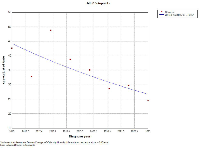
Trend of Abnormal Findings in Eye Disease Screening of Full-Term Neonates from 2016 to 2023.
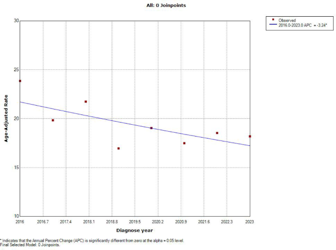
Trend of Retinal Hemorrhage in Full-Term Neonates from 2016 to 2023.
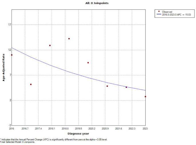
The incidence of retinal white spots showed a downward trend (AAPC = −15%, 95% CI: −29.0% to 1.6%, P = 0.0677, Table 5; Fig. 4), but the trend was not statistically significant. The incidence of retinal exudation showed a downward trend (AAPC = −14.3%, 95% CI: −33.3% to 10.0%, P = 0.2255, Table 5; Fig. 5), with an increasing trend from 2016 to 2020 (APC = 9.3%, 95% CI: −30.9% to 72.9%, P = 0.5818, Table 5; Fig. 5) and a decreasing trend from 2020 to 2023 (APC = −38.1%, 95% CI: −70.0% to 27.9%, P = 0.1260, Table 5; Fig. 5), neither of which was statistically significant. The incidence of retinal FEVR-like lesions showed a downward trend (AAPC = −2.5%, 95% CI: −12.6% to 8.6%, P = 0.5826, Table 5; Fig. 6), but the trend was not statistically significant. The incidence of retinal pigment abnormalities showed a significant downward trend (AAPC = −25.4%, 95% CI: −42.8% to −3.0%, P = 0.0342, Table 5; Fig. 7).
Trend of Retinal White Spots in Full-Term Neonates from 2016 to 2023.
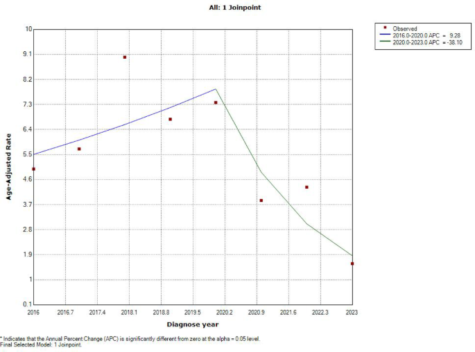
Trend of Retinal Exudates in Full-Term Neonates from 2016 to 2023.
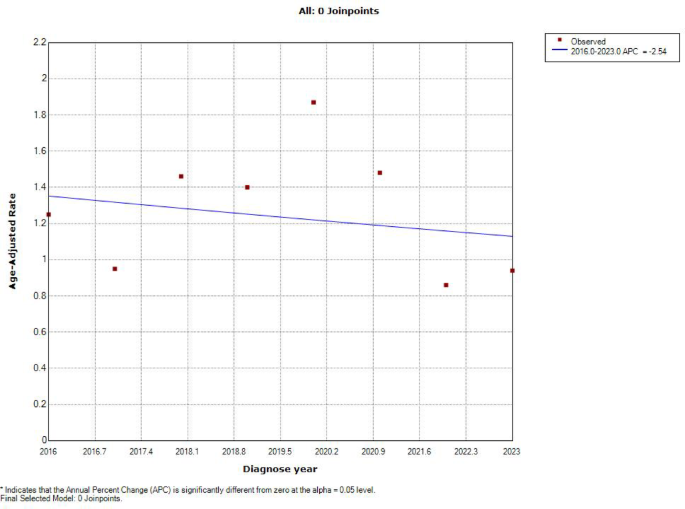
Trend of Retinal FEVR-like Lesions in Full-Term Neonates from 2016 to 2023.
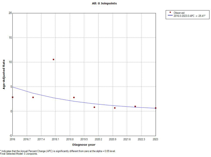
Trend of Retinal Pigment Abnormalities in Full-Term Neonates from 2016 to 2023.
The incidence of other abnormalities showed an upward trend (AAPC = 0.8%, 95% CI: −7.3% to 9.6%, P = 0.8271, Table 5; Fig. 8), but the trend was not statistically significant.
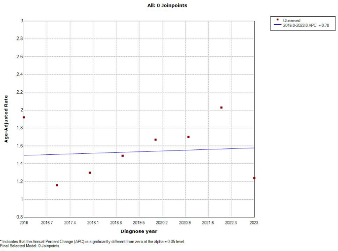
Trend of Other Retinal Abnormalities in Full-Term Neonates from 2016 to 2023.
link